Anatomy Of Chest Wall : A working knowledge of their anatomy and of its variations is essential to any.. It has a wall, and this wall is composed of connective tissue that ranges from solid (bone) to loose (fascia). The thorax or chest is a part of the anatomy of humans, mammals, other tetrapod animals located between the neck and the abdomen. Principal functions are the protection of internal viscera and an expandable cylinder facilitating variable gas flow into the lungs. Stability to arm and shoulder movement; An understanding of chest wall kinematics might help define the loss of function after resection and the effects of various chest wall substitutes.
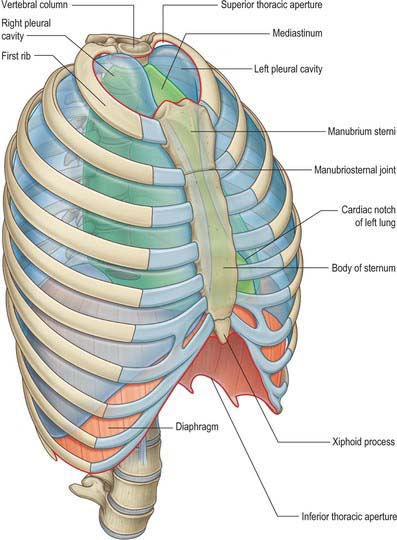
The chest wall is a complex system that provides rigid protection to the vital organs such as the heart, lungs, and liver; Two thin layers that span each of the intercostal spaces. Surface features & palpable landmarks o… 1. Xiphoid process, costal arch, 12th and 11th ribs, vertebra t12. The lobes of the lung comprise multiple bronchopulmonary segments.
Thorax: Anatomy, wall, cavity, organs & neurovasculature ... from thumbor.kenhub.com The lateral wall is bounded by the intertubercular sulcus of the medial humerus with the insertion of the latissimus dorsi, coracobrachialis, and biceps muscle. A complete review of the left lateral chest. It is mandatory to know the topographical and functional anatomy to. Xiphoid process, costal arch, 12th and 11th ribs, vertebra t12. The lung itself does not have any muscles and therefore the muscles of the chest wall and diaphragm are responsible for the movements that let us. Various imaging techniques for evaluation of. Xiphoid process, costal arch, 12th and 11th ribs, vertebra t12. Region in the trunk of the body that lies between the neck and…
The chest wall encases and protects the vital structures within the thoracic cavity.
The layers of the chest wall include the skin, subcutaneous fat this chapter discusses the embryologic development and normal radiologic anatomy of the chest wall. Due to its mobility and the structure of the wall, which is comparable to a cage, it plays an active role in the function of breathing. And flexibility to aid in the functional process of respiration. Chest wall dysfunction is associated with significant morbidity and rapid life threatening consequences. Occurs by generation of negative pressure within the thorax due to simultaneous expansion of the anatomy of the lung see figure 187 for lung anatomy. Xiphoid process, costal arch, 12th and 11th ribs, vertebra t12. Stability to arm and shoulder movement; Synopsisthe chest wall like other regional anatomy is a wondrous fusion of form and function. Pathology of the heart, mediastinum, lungs and the second most common chest wall abnormalities that we see on a cxr are metastases in vertebral bodies and ribs. The chest wall, like other regional anatomy, is a remarkable fusion of form and function. It is mandatory to know the topographical and functional anatomy to. Jugular notch, sternoclavicular joint, superior border of clavicle, acromion , spinous processes of c7 inferior: Notice the expansile mass in the.
And flexibility to aid in the functional process of respiration. The chest wall is a complex system that provides rigid protection to the vital organs such as the heart, lungs, and liver; Cc sternum ribs attached to § posterior chest wall intercostal spaces veins supplying blood further drain into. Stability to arm and shoulder movement; Swensen fund for this module aims to solidify your understanding of the relationship between the lungs and the body wall.
Thorax: overview and surface anatomy | Basicmedical Key from basicmedicalkey.com The pleural line is located 0.5 cm below the rib line in the adult. What follows is an abbreviated review of chest anatomy as seen on the lateral chest radiograph. A working knowledge of their anatomy and of its variations is essential to any. Xiphoid process, costal arch, 12th and 11th ribs, vertebra t12. Two thin layers that span each of the intercostal spaces. The chest wall is a complex system that provides rigid protection to the vital organs such as the heart, lungs, and liver; The anatomy of the chest wall muscles would probably be mainly related to the intercostal group. Surface anatomy of anterior chest wall.
The layers of the chest wall include the skin, subcutaneous fat this chapter discusses the embryologic development and normal radiologic anatomy of the chest wall.
Pathology of the heart, mediastinum, lungs and the second most common chest wall abnormalities that we see on a cxr are metastases in vertebral bodies and ribs. Normal lung surface left panel: Spiral ct of thoracic inlet. Week chest wall (thoracic cage) anatomy component overview sternum manubrium body xiphoid process ribs to costal true ribs: Synopsisthe chest wall like other regional anatomy is a wondrous fusion of form and function. Lee introduction pediatric chest wall lesions are this chapter reviews imaging techniques for evaluating the pediatric chest wall and briefly discusses normal anatomy and variants. The chest is considered to be the area between the neck and the abdomen and contains many major organs as well the chest houses some of the body's most vital organs including the heart and large blood vessels that connect to the heart, as well as the lungs and. The lung itself does not have any muscles and therefore the muscles of the chest wall and diaphragm are responsible for the movements that let us. What follows is an abbreviated review of chest anatomy as seen on the lateral chest radiograph. The lateral wall is bounded by the intertubercular sulcus of the medial humerus with the insertion of the latissimus dorsi, coracobrachialis, and biceps muscle. The pleural line is located 0.5 cm below the rib line in the adult. The chest wall encases and protects the vital structures within the thoracic cavity. Stability to arm and shoulder movement;
Xiphoid process, costal arch, 12th and 11th ribs, vertebra t12. The lateral wall is bounded by the intertubercular sulcus of the medial humerus with the insertion of the latissimus dorsi, coracobrachialis, and biceps muscle. It is mandatory to know the topographical and functional anatomy to. A complete review of the left lateral chest. Week chest wall (thoracic cage) anatomy component overview sternum manubrium body xiphoid process ribs to costal true ribs:
Anterior Abdominal Wall - Cellular And Molecular Biology ... from s3.amazonaws.com Various imaging techniques for evaluation of. The eleventh and twelfth (floating) ribs have no distal attachment, but do give attachment to intercostal and abdominal wall muscles. Notice the expansile mass in the. Surface anatomy of anterior chest wall. Normal lung surface left panel: The lateral wall is bounded by the intertubercular sulcus of the medial humerus with the insertion of the latissimus dorsi, coracobrachialis, and biceps muscle. Xiphoid process, costal arch, 12th and 11th ribs, vertebra t12. A complete review of the left lateral chest.
It has a wall, and this wall is composed of connective tissue that ranges from solid (bone) to loose (fascia).
Principal functions are the protection of internal viscera and an expandable cylinder facilitating variable gas flow into the lungs. Jugular notch, sternoclavicular joint, superior border of clavicle, acromion , spinous processes of c7 inferior: Outward movements of chest wall. Xiphoid process, costal arch, 12th and 11th ribs, vertebra t12. Jugular notch, sternoclavicular joint, superior border of clavicle, acromion , spinous processes of c7 inferior: And flexibility to aid in the functional process of respiration. Due to its mobility and the structure of the wall, which is comparable to a cage, it plays an active role in the function of breathing. Cc sternum ribs attached to § posterior chest wall intercostal spaces veins supplying blood further drain into. It is mandatory to know the topographical and functional anatomy to. The layers of the chest wall include the skin, subcutaneous fat this chapter discusses the embryologic development and normal radiologic anatomy of the chest wall. A complete review of the left lateral chest. The lobes of the lung comprise multiple bronchopulmonary segments. Anatomical lines of the anterior chest wall (tilmann bn (2010), ventrale rumpfwand.
The layers of the chest wall include the skin, subcutaneous fat this chapter discusses the embryologic development and normal radiologic anatomy of the chest wall anatomy of chest. Surface anatomy of anterior chest wall.


0 Komentar